 |
Physicians are treating burn injuries much differently than they did even a decade ago. Though most burn injuries are relatively minor, about 20,000 of the injuries are seen in hospitals with special capabilities in the treatment of burns.
PSP spoke with board-certified plastic surgeon Louis H. Riina, MD, the Director of Wound Care and Hyperbaric Medicine and Assistant Director of the Firefighters’ Burn Center at Nassau University Medical Center on New York’s Long Island. He is also a partner at Long Island Plastic Surgical Group in Garden City, NY.
PSP: What types of burns do you treat?
Riina: We treat all types of burns and exfoliative disorders. All patients with thermal injury are referred into our burn and wound care center by private medical doctors, surrounding hospital emergency medicine departments, outpatient clinics, and by emergency medical services.
We see thermal injuries involving contacts, scalds, flame, as well as chemical-related burns. In addition, our resources are utilized in treating patients with exfoliative disorders such as toxic epidermal necrolysis, Stevens-Johnson syndrome, and patients with large or complex wounds.
Our center sees patients with very small body-surface-area burns and those with very large body-surface-area burns as well. We try to manage burns on an outpatient basis whenever possible, but obviously, larger surface area burns and burns requiring surgical intervention require hospitalization in our burn center.
PSP: How do you treat more superficial burns?
Riina: The initial evaluation that is performed in the clinic or emergency department starts with an appreciation of the size and depth of the injured area. If an injury is partial thickness and there is remaining dermis that is adequate to regenerate upper layers of the skin within a 2-week period, then it is left for the following type of management.
Our outpatient management of burns is novel because we make use of techniques that minimize dressing changes, therefore reducing the discomfort associated with burn care. These techniques allow the body’s healing processes to take place unimpeded by unnecessary interventions.
For example, if a young child sustains a scald injury to his chest by pulling a hot drink off a tabletop, traditionally we would cleanse the wound daily and redress it. Today, we provide a thorough cleansing at the first hospital visit and apply a sustained-release antimicrobial dressing. If that initial dressing becomes adherent to the burned area, much like a scab, it is left alone and intact.
The dressing is antimicrobial—it protects against infection and protects the burned area from contact with the air and is therefore very, very comfortable.
If the burn is mid-depth to superficial, the natural history will be such that the dressing will separate as the skin beneath it heals. A burn treated in this manner will become dry, and the dressing will become adherent to it.
As the skin grows back underneath, the dressing will separate from the healed skin. Usually, these types of burns are resolved in the first week or two.
We do a tremendous amount of outpatient care at our burn center. Our new technique has minimized any unnecessary debridement of healthy tissue. It has been very rewarding for us and very beneficial for the patients as well. This process is very new. I have presented papers on this type of management for several years at the John A. Boswick, MD Burn and Wound Care Symposium and at American Burn Association annual meetings, among others.
PSP: What about more serious, deeper burns?
Riina: Burns that are not expected to heal within a 2-week period are recognized to have a better functional and cosmetic outcome if skin grafted. Our new surgical techniques to treat burns minimize the discomfort for the patient, but also help to improve the reconstructive result.
We affix our skin grafts by using a combination of different glues. We use a fibrin glue to fasten the graft to the recipient bed, and then we use a skin glue to secure the edges of the graft to the healthy skin surrounding it. This results in a minimum of discomfort to the patient, as there are much fewer staples to remove.
These advances in skin graft placement have allowed us to use larger sheet grafts than previously, and the outcome is an overall better cosmetic appearance of the skin grafts as they heal. We pay meticulous attention to detail in the operative care of the patient.
 |
 |
 |
 |
| Figure 1. A 50-year-old male with a scald injury to the dorsum of the foot and ankle—a deep partial thickness to full thickness injury. | |
PSP: Explain, in a little more detail, the newer treatments you are using.
Riina: We use fibrin sealants or glues to affix our skin grafts to the recipient beds. For 10 years now, we have been working on utilizing glues to secure skin grafts in place, as opposed to the traditional sutures or staples. The shortcomings of the previous standard of care, which was suture or staple fixation, are that staples have to be removed and that placement of sutures is time-consuming.
The evolution and refinement of the technique to use fibrin glues to affix skin grafts has been very satisfying for us. It results in the ability to secure the entire length and breadth of the skin graft to the recipient bed, as opposed to the previous standard of care, which only offered point fixation.
This translates to better graft take, better vascularity of the graft, and the ability for us to mobilize the grafted area sooner than we were previously able.
Our topical management of burn injuries differs from the traditional means. The concept of placement of long-term antimicrobial dressings—and then the serial trimming of those dressings as the wound heals beneath them—is new.
The previous standard of care involved removing dressings daily, cleansing and scrubbing the burn injury, and using a topical antimicrobial cream. This was repeated daily until the burn was found to be healed. As you can imagine, the scrubbing of the burn wound is a very painful process, as is the dressing change. That is why, historically, we managed a good portion of these injuries as inpatients.
As we have evolved this “expectant management technique” for partial-thickness injuries, we have been able to do a much larger percentage of this burn care on an outpatient basis.
Children, we find, eat and sleep better when they are at home as compared to when they are in the hospital. Adult patients are much more comfortable in familiar surroundings as well. Since there are no dressing changes to perform, there is no reason patients can’t recuperate at home.
We have been very satisfied with our ability to take care of these partial-thickness injuries on an outpatient basis. And we see the advantages to the patient being able to recuperate at home with a much lower level of discomfort and without unnecessary microdebridement that may be caused by the daily scrubbing of the burn.
We are able to then reserve the excision and grafting procedure for those wounds that either don’t epithelialize in 2 weeks’ time or are immediately recognized as full-thickness or very deep partial-thickness injuries that would be best cared for with early excision and grafting.
In those areas of partial-thickness burns that may have a thin eschar on them, debridement is accomplished by doing serial dressing changes using a nonadherent, antimicrobial dressing. The current one we use is a 100% sodium carboxymethylcellulose dressing with ionic silver.
For those patients whose wounds require surgical reconstruction, we use fibrin sealants or glues to affix our skin grafts to the recipient beds. For 10 years now, we have been working on utilizing glues to secure skin grafts in place, as opposed to the traditional sutures or staples. The shortcomings of the previous standard of care, which was suture or staple fixation, are that staples have to be removed and that placement of sutures is time-consuming.
The evolution and refinement of the technique to use fibrin glues to affix skin grafts has been very satisfying for us. It results in the ability to secure the entire length and breadth of the skin graft to the recipient bed, as opposed to the previous standard of care, which only offered point fixation. This translates to better graft take, better vascularity of the graft, and the ability for us to mobilize the grafted area sooner than we were previously able.
We often combine new technologies with existing techniques, to the patient’s advantage.
To illustrate, a patient may be admitted with an axillary burn and have whirlpool treatment and placement of a topical dressing, go to the operating room, and have the burn excised. Then fibrin sealant will be used to affix the skin graft to that difficult area that the axilla represents, because it has so much of a contour irregularity to which the graft has to conform.
The fibrin sealant allows the skin graft to be anchored throughout that topography. We may also utilize a negative-pressure device to actually suction the graft and dressing into that contoured area.
Here we have two new technologies, the negative pressure or “Vac dressing” and the fibrin sealant for graft fixation, coupled with tried-and-true methods, such as excision, grafting, and hydrotherapy.
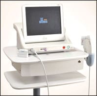
Finally, to mature the scars and to make them more supple, as well as to help remove some of the redness, we currently use a cold laser therapy, which is performed weekly. That therapy is done in conjunction with topical agents to more rapidly mature the scars.
 |
 |
 |
 |
| Figure 2. Five-week-old female with a scald injury to the distal lower extremity and dorsum of foot. This sequence follows the wound through various dressings until it is completely reepithelialized. | |
PSP: What else should we keep in mind with burn treatment?
Riina: Either a burn is going to be monitored and allowed to heal on its own or it is going to have an intervention and require surgical reconstruction. The surgical reconstructions are patient-dependent. We may do one thing on a 6-month-old and something different on a 30-year-old, and something completely different on a 90-year-old. All of [them] have sustained the same injury.
When it comes to taking care of patients surgically, we pay a great deal of attention to blood loss, and we employ various methods of limiting blood loss during the burn excision.
Blood conservation is very important on both ends of the age spectrum. For example, when caring for very young children, we will take steps to minimize the blood loss from the donor site by injecting vasoconstrictive agents in the area from where we are harvesting it.
We will do our excisions under tourniquet to limit the blood loss to the area that is receiving the graft. And we will weigh each of the laparotomy lap pads that are employed during the procedure, so that we are able to know precisely how much blood has been lost by that patient during the surgical process. We can then assess how much blood has been lost and compare it to the circulating blood volume and determine whether that patient should have a cc-for-cc transfusion or whether the patient can tolerate that amount of blood loss without a transfusion.
Conversely, when dealing with very elderly patients, we may accelerate the excision and grafting process.
When we bring an elderly patient to the operating room, [we] try to remove all of the burn and achieve coverage very quickly after the time of injury, so that he or she is recuperating instead of dealing with the burn. They are actually healing instead of allowing the burn and co-morbidities associated with hospitalizing an elderly person to become manifest.
PSP: Tell me about one of your most complex cases.
Riina: One case that comes to mind was the case of a woman who was cooking dinner at her boyfriend’s apartment. She was preparing a pasta dinner with a pot of hot water boiling on the stove. She tripped over the open oven door and the entire stove flipped over and spilled boiling water and boiling pasta sauce onto her right arm and right breast. The wounds were full thickness and required excision and grafting. I had to decide whether to graft onto her excised breast and with which type of graft [to use for the] reconstruction.
I performed her reconstruction using sheet grafts affixed with fibrin sealant, even though they would be the largest ones that I had placed to date, were on an area of irregular topography, and were in a cosmetically sensitive location. Her surgery went as planned, and she had 100% graft take and an excellent cosmetic and functional result.
|
See also “Facial Burns” by Sigrid Blome-Eberwein, MD, in the January 2006 issue of PSP. |
 |
Another memorable case was one where a patient sustained an IV extravisation with an acidic liquid during treatment for metabolic alkalosis. The tissues were necrotic to the level of the biceps muscle. It was this case that led me to the decision to individually rebuild each tissue layer that had been injured. This plan was formulated in order to achieve a reconstruction that did not have a contour irregularity or was tethered to the injured muscle.
I replaced the subcutaneous tissue layer with granulation tissue created with negative-pressure therapy, replaced the dermis with a dermal regeneration template, and replaced the epidermis with a thin epidermal autograft. The epidermal and dermal tissues were affixed with fibrin sealant. This case is a perfect example of existing techniques and new technology being utilized together to great patient advantage.
The reconstruction was accomplished without contour deformity and with an excellent cosmetic and functional result.
Amy Di Leo is a contributing writer for PSP. She can be reached at [email protected].


